Rasmussen’s Encephalitis: A Rollercoaster Ride
Hello to everyone over there on the big island from those of us who work here on the little island, Tassie. Let me quickly introduce myself, I commenced working in the field of clinical neurophysiology back in 1993. Back when brainwaves were recorded upon analogue systems nearly the same length as a patient trolley! Yes, I have seen some wonderful, computerised advances during my career. Not so wonderful though are the neurological mysteries, the lack of understanding of their cause, the resulting dilemmas in clinical decision making and the roller-coaster of a ride that families must endure. Rasmussen’s encephalitis (RE) is one such neurological mystery I briefly touch upon here and the (EEG) journey of one of our young patients.
Let me first start by explaining that there simply isn’t enough “blogging” space to entirely cover RE.
So, what is RE? First described by Neurosurgeon Theodore Rasmussen in the late 1950s, RE is a rare progressive disease mostly affecting children or young adults. RE is characterised by unilateral encephalitis of one cerebral hemisphere, drug resistant epilepsy and progressive chronic neurological and cognitive deterioration. Management of the disease is confounded by difficulties in making an early diagnosis, a lack of understanding of the cause, and the varying clinical features that create quandaries and catch-22s in clinical decision making. There is growing evidence for an immunopathalogical basis of RE, and the main players for CNS degeneration can be tagged into three types: antibody-mediated CNS degeneration, T-cell cytotoxicity, and microglia-induced degeneration. However, the primary cause remains unknown.
Patients with RE undergo several diagnostic procedures. EEGs show widely varying abnormalities related to clinical progression as the disease process affects different parts of the brain at different times. MRI and PET are essential investigations for focal cortical atrophy and hypometabolism. CSF will show evidence of CNS inflammation. IV methylprednisolone is administered to slow the progression of the disease but is not a cure. Seizures will be refractory to trials of several antiseizure medications. The only cure for RE is either functional hemispherectomy surgery (disconnection of the affected hemisphere) or anatomical hemispherectomy (removal of the diseased hemisphere).
Mid 2019 we meet a 3yo with new onset seizures (GTCS and absence within 1 month), past history of febrile seizure at 18 months, no previous EEG, and normal development. An urgent EEG is performed which shows irregular delta and theta frequencies over the right hemisphere, and during HV, focal right sided spike-wave discharges are seen maximally in the right frontal region. MRI reveals a subtle right sided malformation of cortical development. Treated with TPM and LEV.
Soon our patient presents with frequent focal seizures. Urgently admitted for overnight monitoring to investigate frequent focal aware seizures with insular-opercular features, left facial jerking and prominent salivation, along with separate episodes with left forearm sensory changes. Medications TPM, LEV, LTG are commenced. The awake and asleep interictal EEG shows multifocal right-sided polyspike-wave, spike-wave and sharp-wave discharges maximal over the right frontal region. No clinical events are captured. Further imaging shows subtle increase in the extra-axial spaces over the right hemisphere and PET shows diffuse hypometabolism through the right frontotemporal region. A diagnosis of RE is made. A second overnight monitoring admission is organised to capture events. The patient is having multiple daily focal seizures with left version, left arm stiffness, left facial jerking and prominent salivation. Medications are LEV, CLB, LTG (to switch to Lacosamide). Finally, an electroclinical seizure with left versive features is captured with an early right central-frontocentral ictal rhythm along with myoclonic jerks in the awake and asleep states. There is further electroclinical localisation of focal epileptiform activity to the right central-frontocentral region.
EEG snippets from the second overnight VEM (double-banana montage):
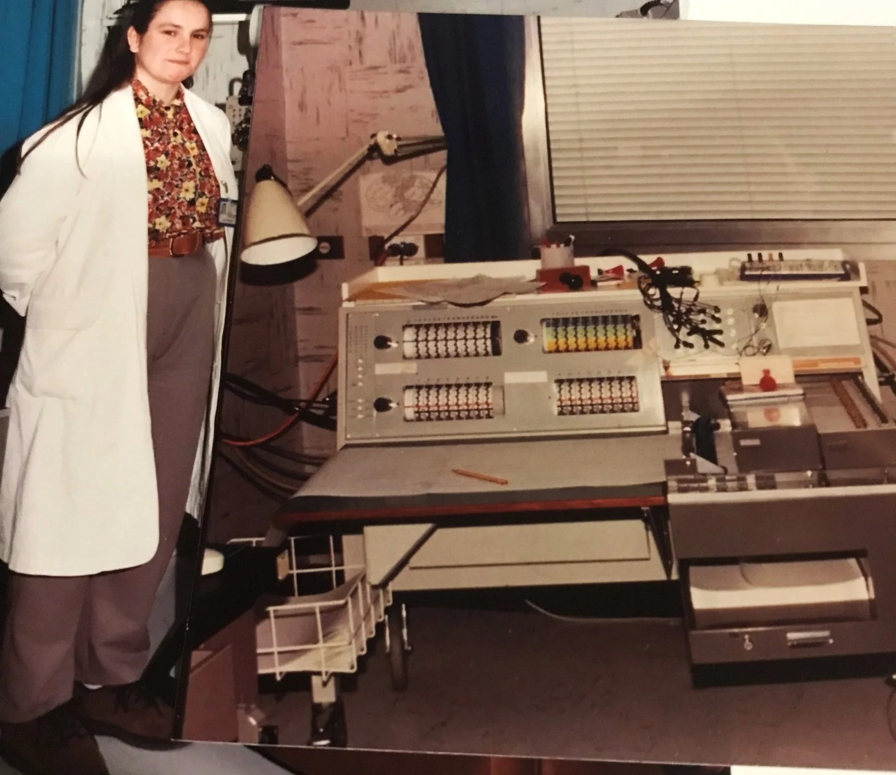
Kim as a trainee in early 1990s with a Mingograph.

Kim Wilmot
Senior Neurophysiology Scientist at Royal Hobart Hospital

Myoclonic Jerk

Seizure: forced left version of the head and eyes, then left arm flexion
Seizure frequency and severity continues to escalate. Four-weekly induction pulse corticosteroid IVIG is commenced. Our patient is soon referred to the mainland for neurosurgery, a right functional hemispherectomy. The functional hemispherectomy/commissural disconnection for RE involved a resection of the posterior frontal, insular and anterior right temporal lobe, right cingulate gyrus and right side of the corpus callosum. Left hemiplegia, acquired brain injury and visual neglect were all expected following the surgery. The surgery resulted in complete clinical seizure freedom while being off all antiseizure medications, although with severe persistent neurological impairments (as predicted).
Fast forward to February 2023, when the patient is discharged from Paediatric neurology (with continued follow-up with general paediatrics) as clinically seizure free and off medication since Nov 2020. What a wonderful outcome for patient and family!
Then….
May 2023 our patient begins to experience multiple daily episodes of persistent right sided headache with periods of severe exacerbations that last on and off for hours, along with nausea and phonophobia. A 2-hour EEG is performed whereby 27 rolling electrographic seizures are recorded over the disconnected right hemisphere. Many electrographic seizures have a temporal correlation to symptoms and button presses (right head pain). There are also electrographic seizures which occur without button presses. There are no demonstrated motor manifestations of the electrographic seizures, in particular no version or hemiclonic jerking. There is no left hemisphere interictal abnormality or progression of the right electrographic seizures to involve the left hemisphere. Our patient is diagnosed with “status migrainosus”: a consequence of electrographic epilepsia partialis continua in the disconnected right hemisphere and presumed to be a manifestation of the ongoing encephalitis.
Four weeks later, multiple daily episodes of head pain continue with the patient now on Topiramate. A follow up 3-hour EEG shows 55 rolling electrographic seizures in varying right hemisphere regions.
EEG snippets (Source Derivation) below:

Maximal discharges T4-T6 during button press (head pain).

Maximal discharges Fp2 with no button press.
Two weeks later, pulse IV methylprednisolone is given along with Topiramate, daily aspirin and neurofen. Follow up 2-hour EEG in July reveals 5 electrographic seizures with varying right hemisphere regions of onset and evolution. No headache events are recorded. The EEG pattern and headaches initially subsided with Pulse IV methylprednisolone.
Follow up 2-hour EEG in August (now ~ 3 months after initial presentation of head pain and after second course of Pulse IV methylprednisolone) shows 25 rolling electrographic seizures arising from the disconnected right hemisphere, with regions of onset and evolutions involving posterior, central, temporal and frontal regions. Seizure durations vary between 50sec-120sec. None of the electrographic seizures have a clear clinical correlate and there were no event button presses (i.e., no head pain events), however parents stated “it’s a good day today.” There is no left hemisphere interictal abnormality.
Our patient continues to experience recurrent bouts of severe headache despite daily aspirin and neurofen, plus Topiramate. Further MRI shows that the right basal ganglia and thalamus appear to be smaller, but no active inflammation seen, and the left hemisphere is normal.
Post-functional hemispherectomy patients can have EEGs showing electrical status in the disconnected hemisphere, alternating IEDs and subclinical seizures, presenting as a migraine-like syndrome. Migraines continue for weeks to months and tend to not respond to antiseizure medication and immunotherapies. Anatomical hemispherectomy is now being considered by family.
A future blog topic…
Consent for this blog kindly given by family.
References
1. Sophia Varadkar, et al. Rasmussen’s encephalitis: clinical features, pathobiology, and treatment advances. Lancet Neurol. 2014 February: 13(2): 195-205
2. Emma Macdonald-Laurs, et al. Clinical seizure manifestations in the absence of synaptic connections. Epileptic Disord 2021: 23 (1): 167-172